Home / Microfluidic Resources / Microfluidic Reviews / Solvent-Mediated Microfluidic Generation of Cell-Sized Liposomes
Microfluidic research summary

Published on 06 May 2026

Solvent-Mediated Microfluidic Generation of Cell-Sized Liposomes
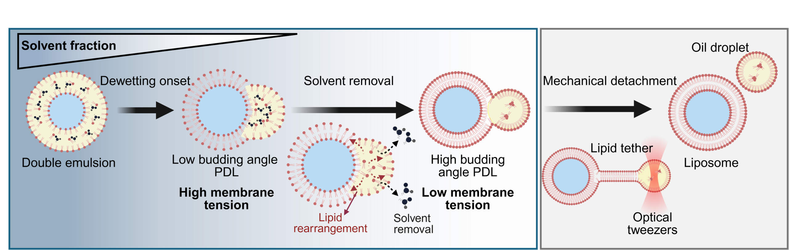
Table of Contents

Solvent-Mediated Microfluidic Generation of Cell-Sized Liposomes

Graphical abstract DEs

This Research Summary article is based on the papers titled “Facile and versatile PDMS-glass capillary double emulsion formation device coupled with rapid purification toward microfluidic giant liposome generation,” authored by Bakouei M, Kalantarifard A, Sundara Raju I, Avsievich T, Rannaste L, Kreivi M, Elbuken C., and “Solvent-Mediated Dewetting Principles for Cell-Sized Liposome Formation,” authored by Bakouei, M.†, Avsievich, T.†, Sundara Raju, I., Stumpf, D., Kalantarifard, A., Ryplida, B., & Elbuken, C.

Introduction to Cell-sized Liposome

The reconstitution of cell-sized liposomes as fundamental building blocks for synthetic cells is of critical importance with far-reaching applications in cell biology, biomanufacturing, and therapeutics. Microfluidic technologies enable precise control over the formation and composition of these synthetic cells, offering throughput and monodispersity superior to conventional generation methods. However, microfluidic liposome production is often hindered by fabrication complexity and the intricate dynamics of the generation process. Here, the team demonstrate a hybrid microfluidic double emulsion formation chip and a solvent-mediated dewetting method aimed to streamline liposome generation for application in synthetic biology. Furthermore, this approach explains dewetting-based, cell-sized liposome assembly.
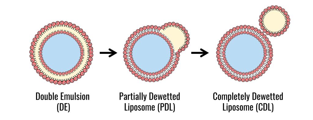
double emulsion dewetting steps

Double emulsion for liposome generation Setup

Materials

  • Microfluidic hybrid formation chip (Produced via microfabrication using 3D printer mold, Form 3+ Formlabs and standard soft lithography)
  • DMIL LED inverted microscope (Leica)
  • FastCam mini high-speed camera (Photron)
  • OB1 MK3+ Pressure Controller (Elveflow)
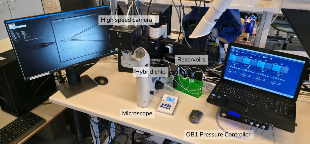
setup for DE generation computer, microscope, chip, pressure controller elveflow
Microfluidic setup for the high throughput generation of cell-sized liposomes

Hybrid microfluidic double emulsion formation chip 

Bakouei et al. developed a PDMS-glass capillary hybrid chip as a treatment-free and versatile approach for generating double emulsions (DEs), offering an alternative to standard PDMS chips and glass-capillary co-flow devices.

This hybrid architecture leverages the inherent surface properties of both PDMS and glass to provide the

  • spatial hydrophobicity and spatial hydrophilicity

required for DE formation (Fig. 1), thereby eliminating the need for intricate surface treatments that suffer from a lack of repeatability and short longevity.
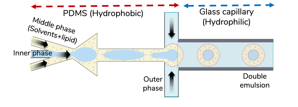
Schematic demonstration of double emulsion (DE) formation using PDMS-Glass capillary hybrid chip
Fig. 1: Schematic demonstration of double emulsion (DE) formation using PDMS-Glass capillary hybrid chip

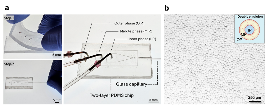
Consequently, the hybrid chip enables DE generation in a simplified, reusable, and versatile manner. Fig. 2a illustrates the two-step assembly of the hybrid chip:

  • two identical patterned PDMS replicas are aligned and bonded
  • a tapered capillary with specific tip dimensions is inserted into the PDMS housing.

The assembled chip features two consecutive flow-focusing junctions.

  • The first junction, situated within the naturally hydrophobic PDMS, facilitates water-in-oil (W/O) droplet formation.
  • The second junction, located within the hydrophilic glass capillary, facilitates the high-throughput formation of water-in-oil-in-water double emulsions (W/O/W) (Fig. 2b).
The 2-step assembly illustration of the chip and photograph of the chip with its corresponding elements b) High yield thin shell DE formation using the hybrid chip
Figure 2. a) The 2-step assembly illustration of the chip and photograph of the chip with its corresponding elements b) High yield thin shell DE formation using the hybrid chip

Inlet tip diameter influences the size of the double emulsion

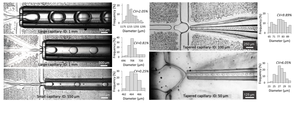
As shown in Fig. 3 and Movie 1, by modulating the inserted capillary tip diameter, a wide size range of monodisperse thin-shell double emulsions can be generated, spanning from 1.2 mm down to 27 µm.
Versatility in the formation of thin shell DEs using the hybrid chip
Fig. 3: Versatility in the formation of thin shell DEs using the hybrid chip
Movie 1: The hybrid chip enables the tunability of the double emulsion size.

Pressure influences the shell thickness

The shell thickness and topology of the DEs can be precisely controlled by modulating the pressures applied across the three phases and by selecting specific formation modes. As demonstrated in Movie 2, the hybrid chip functions under three formation modes.

The double-dripping mode is recommended for the high-yield production of Cell-sized thin-shell DEs.

Movie 2: Thin shell double emulsion formation modes using the hybrid chip.

Key Findings

Solvent-mediated dewetting transition from double emulsion to partially dewetted liposome

The team elucidates that the dewetting transition from microfluidically generated DEs to liposomes is driven by solvent removal. DEs were generated using a mixture of 60%:40% combination of hexanol and paraffin oil as the solvents of DOPC lipids in the middle phase (MP). Upon formation, the hexanol, being water-soluble and volatile, starts partitioning out of the DE shell. This solvent removal triggers the spontaneous onset of dewetting, resulting in a partially dewetted liposome (PDL) (Movie 3), which is characterized by the contact angle of its residual oil pockets, referred here as the budding angle.

Movie 3: Onset of double emulsion dewetting resulting in a liposome with an attached oil pocket.

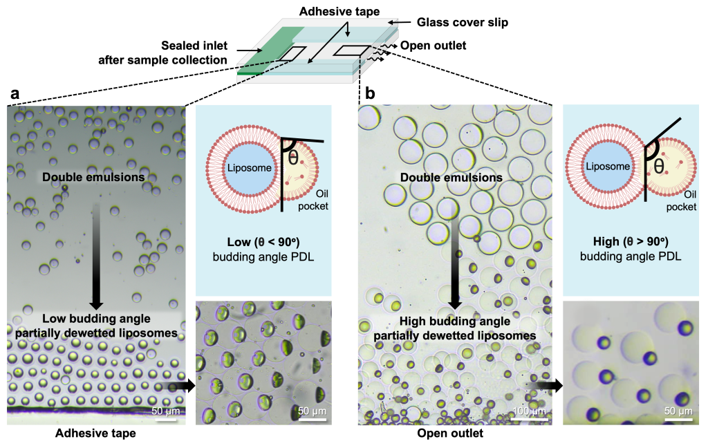
In a semi-permeable glass chamber, solvent removal proceeds extremely slowly, leading to spatial variations in dewetting progression (Fig. 4). DEs situated near air-exposed regions experience a faster dewetting transition, resulting in a high budding angle morphology (θ> 90°). In contrast, those lacking air exposure undergo a slower dewetting transition, resulting in a low-budding-angle morphology (θ < 90°).
Spatially non-uniform partial dewetting transition reveals the regulatory role of solvent removal
Figure 4. Spatially non-uniform partial dewetting transition reveals the regulatory role of solvent removal. a) Dewetting gradient observed near the semipermeable adhesive tape: intact DEs in the central region and partially dewetted liposomes (PDLs) with a low budding angle (θ < 90°) near the semipermeable edges. b) Dewetting gradient near the open outlet: DEs rapidly transition into PDLs with high budding angle.

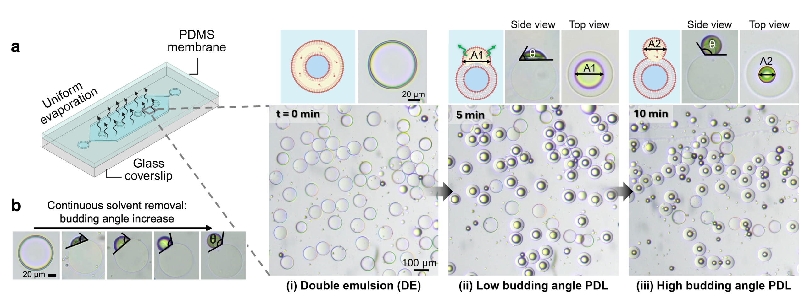
To address the slow and spatially non-uniform dewetting transition, a gas-permeable PDMS chamber was utilized for DE collection. Incorporating a thin PDMS membrane in the chamber ensures a high hexanol removal flux. This approach resulted in a spatially synchronous dewetting transition from DEs to high budding angle PDLs within 10 minutes (Fig. 5 and Movie 4).
Uniform dewetting transition of freshly collected DEs into low budding angle PDLs, and then upon sufficient solvent removal to high budding angle PDLs within a PDMS chamber
Figure 5. Uniform dewetting transition of freshly collected DEs into low budding angle PDLs, and then upon sufficient solvent removal to high budding angle PDLs within a PDMS chamber
Movie 4: Vesicles post solvent removal in a PDMS chamber, demonstrating high budding angle morphology.

Origin of dewetting morphological transformation upon solvent removal

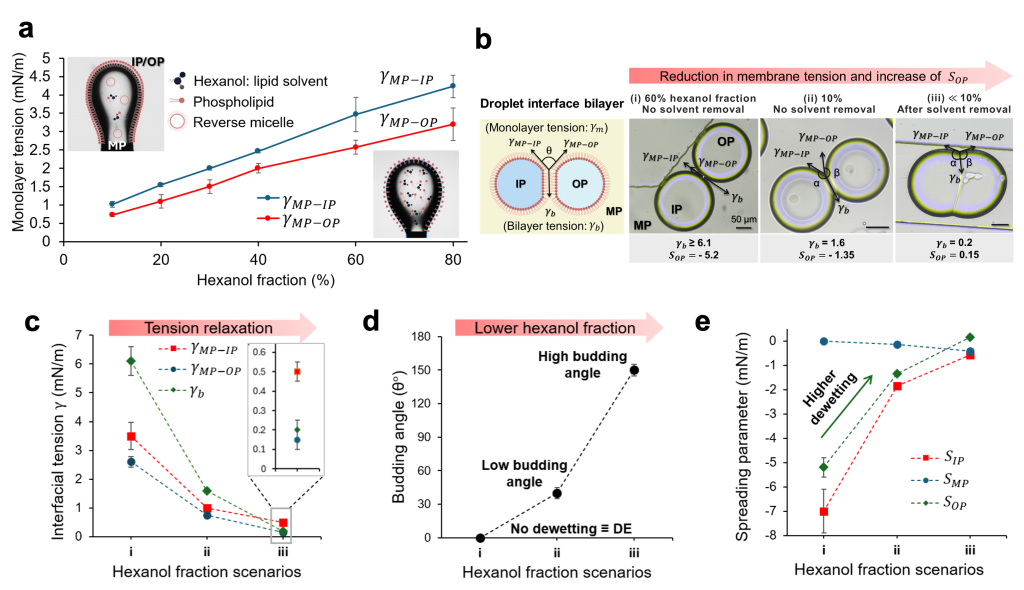
To elucidate the mechanism underlying the dewetting transition from double emulsions (DEs) to high budding angle partially dewetted liposomes (PDLs) via solvent removal, the process within the framework of ternary phase wetting theory was analyzed.

It was hypothesized that solvent removal simultaneously reduces the interfacial tension of both

  • oil-aqueous monolayers (Fig. 6a) and
  • the bilayer membrane (Fig. 6b).

This significant reduction in tension, with the membrane tension decreasing more rapidly than the monolayer tensions (Fig. 6c), establishes a new equilibrium, resulting in an altered dewetting morphology (Fig. 6d).

Consistently, the analysis of the spreading parameters, predictive of wetting morphology, demonstrates that solvent removal increases the outer phase spreading parameter ( ), thereby favoring dewetting transition (Fig. 6e).
Solvent removal results
Figure 6. Solvent removal results in a) Reduction of monolayer tension and b) membrane tensions that drive morphological transitions, favoring dewetting. The measurements were conducted for monolayer tension via tensiometry and for membrane tension via droplet interface bilayer morphometry. Analysis of c) interfacial tensions, d) budding angle, and e) spreading parameters under three hexanol fraction scenarios shown in (b).

Non-spontaneous nature of complete dewetting

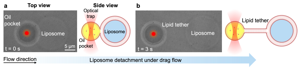
Solvent removal facilitates the morphing of almost fully budded liposomes; however, the liposome and oil pocket maintain a shared neck that keeps them adhered. This suggests that complete detachment is not spontaneous and fission requires an external force.

A characterization of the residual adhesion and the magnitude of the mechanical force required for complete dewetting was then performed. Using optical tweezers, the formation of lipid tethers anchoring the oil pocket was observed, which require a detachment force on the piconewton scale to achieve separation (Fig. 7 and Movie 5).
 An optical tweezers experiment
Figure 7. An optical tweezers experiment demonstrating a partially dewetted liposome under drag force. Continuous drag force exerted by the flow on the trapped PDL pulls against the liposome (a), leading to its separation and the formation of a lipid tether (b).
Movie 5: Detachment of oil pocket from liposome using optical tweezers.

High-yield liposome generation with complete dewetting transition

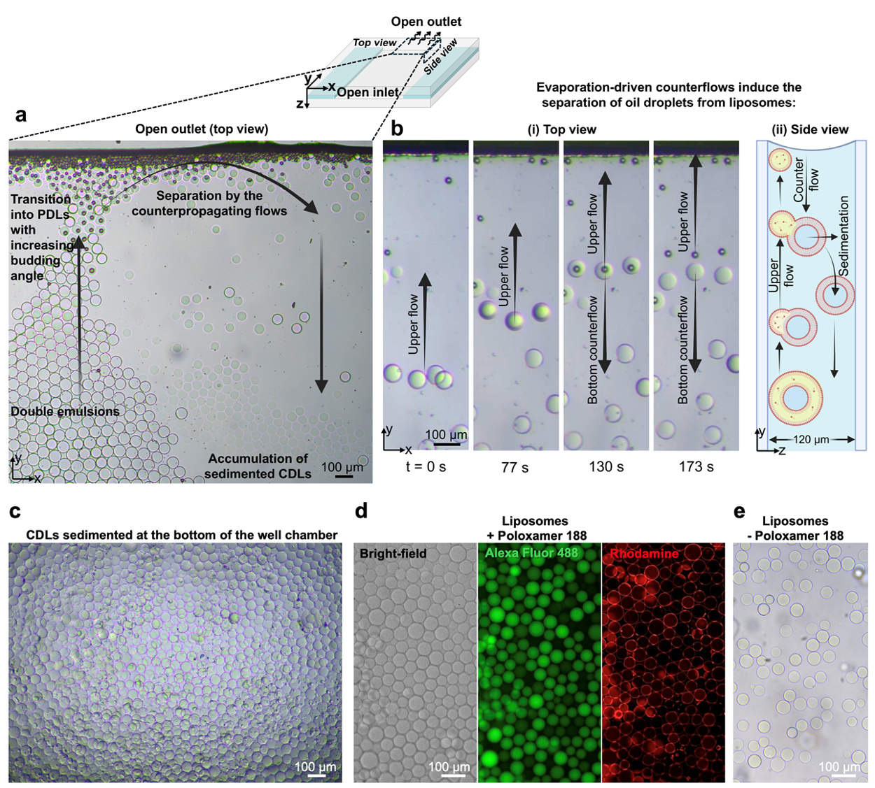
Solvent removal and mechanical force are essential mechanisms for dewetting and liposome formation. Through evaporation-driven flow and buoyancy-induced separation, high-throughput liposome generation was achieved. Complete separation of the liposomes from the oil pockets was enabled by

  • hexanol evaporation and
  • shear flows induced by the evaporation of the bulk outer phase (Fig. 8a,b).

While hexanol depletion promoted the dewetting transition to a high budding angle PDL, concurrent evaporation of the outer phase generated internal flows. Sufficient shear forces were thereby created to enable complete dewetting.

Similarly, efficient formation was observed when DEs were collected into a large open-well chamber immediately after generation (Fig. 8c,d). Hexanol evaporation from DEs floating at the surface was facilitated by the lidless chamber. Once a high-budding-angle morphology was reached, pulling stress was applied by buoyancy forces at the oil pocket–membrane interface, promoting complete dewetting. Additionally, liposomes were formed in the absence of surfactants (Fig. 8e), offering greater biocompatibility, as membrane properties may be adversely affected by surfactants, thereby inhibiting biomolecular interaction with synthetic cells.
Evaporation-driven counterflows enable high-yield complete dewetting of liposomes. b) Time-series images of DEs transition into low and high budding angle PDLs followed by complete detachment of liposomes c) Brightfield and fluorescent images of high-yield CDLs accumulated at the bottom of the well, d) demonstrating encapsulated Alexa Fluor 488 in cargo and Rhodamine in the liposome membrane. (e) Liposomes formed without poloxamer 188 surfactant.
Figure 8. a) Evaporation-driven counterflows enable high-yield complete dewetting of liposomes. b) Time-series images of DEs transition into low and high budding angle PDLs followed by complete detachment of liposomes c) Brightfield and fluorescent images of high-yield CDLs accumulated at the bottom of the well, d) demonstrating encapsulated Alexa Fluor 488 in cargo and Rhodamine in the liposome membrane. (e) Liposomes formed without poloxamer 188 surfactant.

Conclusion

Here, a hybrid formation chip and solvent-mediated dewetting method are offered as a microfluidic cell-sized liposome generation system.

  • Hybrid chip by leveraging PDMS and glass capillary inherent properties enables the treatment-free, versatile, and reusable formation of double emulsion templates.
  • The solvent-mediated dewetting enables morphing of liposomes from these templates through solvent removal and mechanical force mechanisms.

The extent of solvent removal governs the oil droplet-liposome dewetting progression and equilibrium morphology. Solvent removal through alteration in monolayers and membrane tension enforces new equilibrium morphologies, favoring dewetting. This research highlights that completion of solvent removal results in an almost budded oil droplet with an attached necking requiring a pN-scale external force for detachment, identified using optical tweezers. This work demonstrates a predictable and high-throughput approach for microfluidic cell-sized liposomes generation in a biocompatible manner.

Author Information

Caglar Elbuken: Professor at the Faculty of Biochemistry and Molecular Medicine, University of Oulu, and VTT technical research center of Finland, and head of the Microfluidics and Biosensor research group

Mostafa Bakouei: PhD researcher at the Faculty of Biochemistry and Molecular Medicine, University of Oulu


Written and reviewed by Mostafa Bakouei, and Louise Fournier, PhD in Chemistry and Biology Interface. For more content about microfluidics, you can have a look here.

The Best Flow Control Technology

Discover the OB1

What are cell-sized liposomes?

Cell-sized liposomes, also known as giant unilamellar vesicles (GUVs), are synthetic membrane-bound compartments that mimic certain structural features of biological cells. They are widely used in synthetic biology, membrane biophysics, drug delivery research, and artificial cell development.

Why are microfluidics used to generate liposomes?

Microfluidics provides precise control over fluid flows, enabling the production of highly monodisperse liposomes with controlled size, composition, and encapsulation properties. Compared to conventional bulk methods, microfluidics offers better reproducibility and higher throughput.

What is the principle behind this liposome generation method?

The method relies on the formation of water-in-oil-in-water (W/O/W) double emulsions, which serve as templates for liposome formation. Following double emulsion generation, a solvent-mediated dewetting process removes the oil layer, leaving behind a lipid bilayer membrane that forms the liposome.

What is a double emulsion?

A double emulsion is a droplet encapsulated inside another droplet. In this work, an aqueous core is surrounded by a lipid-containing oil shell, which is itself dispersed in an outer aqueous phase. These structures act as intermediates during liposome formation.

What is unique about the PDMS-glass hybrid microfluidic chip?

The chip combines the natural hydrophobicity of PDMS with the natural hydrophilicity of glass. This design eliminates the need for complex surface treatments typically required for double emulsion formation, resulting in a simpler, more robust, and reusable device.

How does the hybrid chip generate double emulsions?

The device contains two consecutive flow-focusing junctions:

  1. A hydrophobic PDMS junction forms water-in-oil droplets.
  2. A hydrophilic glass capillary junction encapsulates these droplets within an outer aqueous phase, creating water-in-oil-in-water double emulsions.

What role does pressure control play?

Pressure control regulates the flow rates of the inner, middle, and outer phases. By adjusting these pressures, researchers can precisely control droplet size, shell thickness, and formation stability. The study used an OB1 MK3+ Flow Controller for this purpose.

How can the size of the double emulsions be controlled?

The size of the double emulsions is primarily adjusted by changing the diameter of the inserted glass capillary tip and tuning the applied pressures. Using different capillaries, the researchers generated droplets ranging from approximately 1.2 mm down to 27 µm.

How is shell thickness controlled?

Shell thickness is controlled by adjusting the pressure applied to the inner aqueous phase and selecting appropriate droplet formation regimes. Higher inner-phase pressures generally produce thinner oil shells, which are desirable for liposome formation.

Which droplet formation mode is recommended for liposome production?

The study identified the double-dripping mode as the preferred operating regime for producing high yields of thin-shell double emulsions suitable for cell-sized liposome generation.

What is solvent-mediated dewetting?

Solvent-mediated dewetting is the process through which the oil shell surrounding the aqueous core is progressively removed. As solvent leaves the oil phase, lipid molecules reorganize into a bilayer membrane, transforming the double emulsion into a liposome.

Is solvent removal alone sufficient to form liposomes?

Solvent removal is sufficient to initiate the transition from double emulsions to partially dewetted liposomes. However, complete dewetting and detachment of residual oil pockets often require additional mechanical forces.

Why is understanding dewetting important?

A mechanistic understanding of dewetting helps researchers optimize liposome production, improve membrane purity, reduce solvent residues, and develop more reliable synthetic cell platforms.

What are the main applications of cell-sized liposomes?

Applications include:

  • Synthetic biology and artificial cells
  • Drug delivery research
  • Membrane biophysics studies
  • Biomanufacturing
  • Encapsulation of biomolecules
  • Studying cellular processes such as budding and compartmentalization

Why was pressure-driven microfluidics chosen for this setup?

Pressure-driven flow control provides stable, pulseless, and highly responsive flow regulation, which is critical for maintaining consistent double emulsion formation and achieving reproducible liposome production.

Bakouei M, Kalantarifard A, Sundara Raju I, Avsievich T, Rannaste L, Kreivi M, Elbuken C. Facile and versatile PDMS-glass capillary double emulsion formation device coupled with rapid purification toward microfluidic giant liposome generation. Microsyst Nanoeng. 2024;10(1):183

Bakouei, M., Avsievich, T., Sundara Raju, I., Stumpf, D., Kalantarifard, A., Ryplida, B., & Elbuken, C. Solvent‐Mediated Dewetting Principles for Cell‐Sized Liposome Formation. Small, 2026;22(18): e12610.

Share the Post:
Contact
How can we help you?
Quoteor technical request
Job application Job
application
Collaboration or partnerships Collaborations
or partnerships
Customer support Customer
support
Others questions Other

For more information or Technical discussion

Contacting for
a job application?
We are happy that you are interested in Elveflow. You can apply to our open jobs or send us your open application. Over here!

For more information or Technical discussion

Need customer support?

For more information or Technical discussion