Highlights
Setup
SPATIAL TRANSCRIPTOMICS SYSTEM SETUP
FLUIDIC SETUP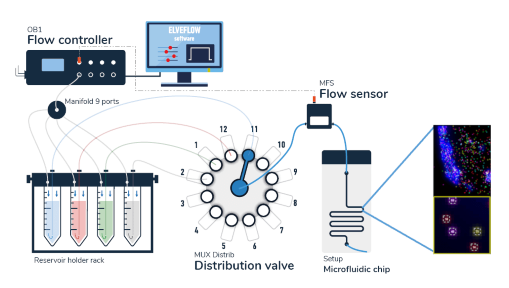
The pack contains:
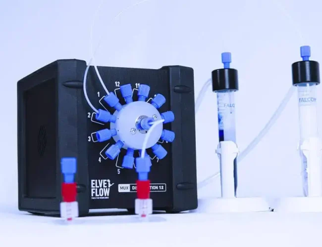
- OB1 flow controller
- Microfluidic flow sensor
- One or two 12:1 MUX distribution valves depending of the number of dyes you want to inject
- Tubings and luers
- Several Eppendorfs or Falcon reservoirs
- Microfluidic chips
- Microfluidic Software
- A user guide
upcoming Microscope Setup
Applications
Why use it?
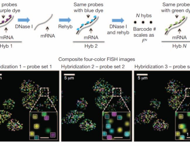
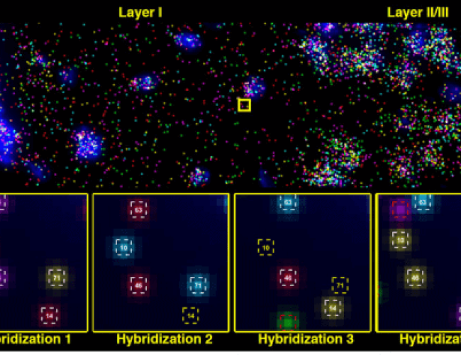
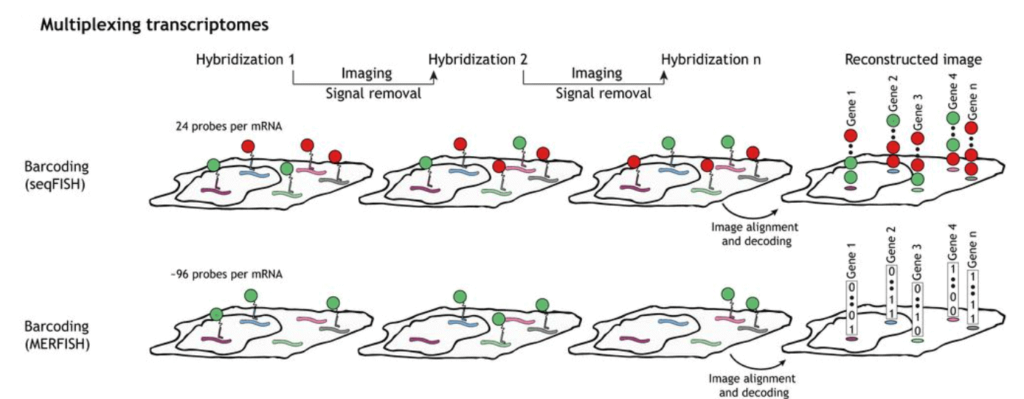
Microfluidics is the most effective way to observe multiple genes and their spatial configuration through MERFISH (Multiplexed Error-Robust Fluorescence In Situ Hybridization) or seqFISH (sequential Fluorescence In Situ Hybridization), as it allows for experimentation with a significantly smaller amount of costly dye and buffer solutions and is compatible with biological applications and microscope observations.
As was already explained, the solutions can be injected into the cell using an automated process. Additionally, the system can be configured to link many chips, making it simple to view multiple samples simultaneously.
Before setting up a fluorescence in situ hybridization system, this pack can also be used in conjunction with other microfluidic procedures; for instance, microfluidic single-cell encapsulation can be used for single-cell isolation [1].
Microfluidics can also be applied to barcoding (DBiT-seq) or the oscillatory flows of diluted probe solutions used in the MA-FISH approach.
Over the course of more than a decade, the Microfluidics Innovation Center acquired competence in microfluidics. It can offer its cutting-edge biological and engineering skills, making it the ideal partner for you as you make the switch to microfluidics.

 Job
Job Collaborations
 Customer
 Other
Other