Microfluidics application note
Published on 27 August 2019
Figure 1: This tissue-engineering ventricle, made from neonatal rat ventricular myocyte tissue, is spontaneously contracting, sutured and attached to a catheter. Credit: Luke MacQueen/Disease Biophysics Group/Harvard SEAS
Until recently, microfluidic devices have been employed to support tissue-engineering experiments on basal lamina, vascular tissue, liver, bone, cartilage and neurons as well as organ-on-chips. The major contribution of microfluidics is with complex tissue morphogenesis, where microfluidic structures ensure a steady blood supply while minimizing experimental volumes and ensuring easy reproducibility. Instead of an in-vitro convective system with discrete conditions, microfluidics combined to tissue-engineering and organ-on-chip offers continuous conditions in transition system between in-vitro and in-vivo models [2].
This abstract covers a research article published in July 2018 in Nature Biomedical Engineering. The WYSS and Boston Hospital created a model of the human left ventricle (1/250 size reduction), made of nanofibrous scaffolds and monitored by catheter sensors. These scaffolds promote native-like anisotropic myocardial tissue genesis and chamber-level contractile function. Using Elveflow’s OB1 Mk3 Flow Controller, they managed to deliver a precise cyclic pressure to the extra-ventricular loop driving intraventricular fluid flow via ventricle contraction.
TISSUE-ENGINEERING APPLICATIONS
- Scale model of the human left heart ventricle (transition between in vitro and in vivo models)
- Preclinical cardiology
- Regenerative medicine research (disease model of structural arrhythmia)
- Drug screening
[ooc_rebound]
METHODS
First, Luke A. MacQueen et al. produced ellipsoidal thin-walled chambers composed of a nanofibrous synthetic–natural polymer–protein blend by pull spinning technique.
Then, chambers were seeded with neonatal rat ventricular myocytes (NRVMs) or hiPSC-Cardiomyocytes. The chambers had enough porosity to support cell infiltration and promote tissue morphogenesis (cf. Figure 2)
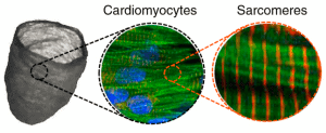
Figure 2 : Schematics of ventricle surface immunostaining (F-actin, DAPI, α-Actinin)
Finally, chambers were sutured to tubing and bioreactor components through which catheter were introduced. These catheters were connected to the Elveflow’s OB1 Mk3 Flow Controller, producing the contraction of both NRVM and hiPSC-CM ventricles (cf. Figure 3).
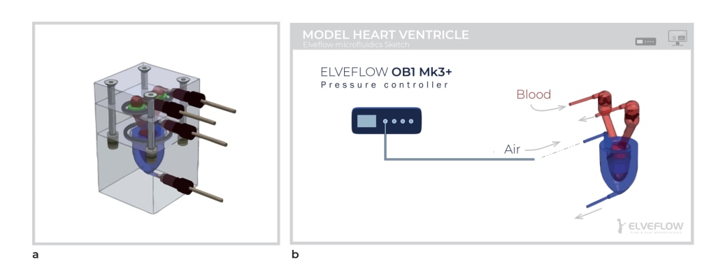
Figure 3 : (a) Heart bioreactor (HBR) computer-aided design. (b) Flow loop schematic.
MICROFLUIDIC RESULTS FOR TISSUE-ENGINEERING
The OB1 MK3 pressure controller enabled the team to generate small amplitude pressure variations (ΔP=0.1mmHg, equal to ΔP=0.13mBar, at 2Hz) applied to the tissue engineered ventricle scaffold through the extra-ventricular channel, driving fluid flow through the intraventricular flow loop.
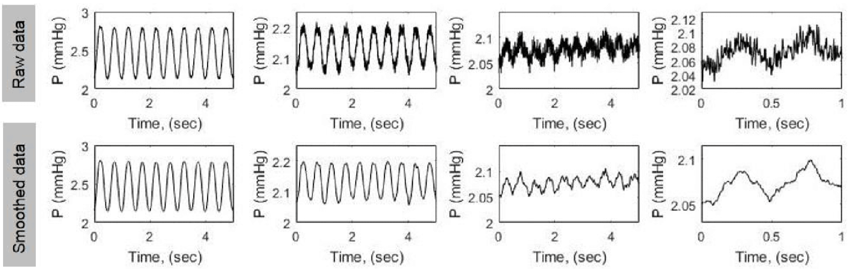
Figure 4 : Small-amplitude pressure recordings obtained by assisted ventricle scaffold contraction using our instrumented bioreactor’s ventricular assist channel and pressure supplied by a programmable pressure-driven microfluidic pump
By generating successively smaller peak-to-peak pressure sinusoids, it was proved that pressure and volume variations generated by their tissue-engineered model ventricles were above the background noise.
CONCLUSION
This article showcases the feasibility of developing a functional scale model of the heart ventricle, using tissue engineering and microfluidics to assist contraction of a nanofibrous ventricle. This work offers great opportunities for multiscale in vitro cardiology assays and regenerative medicine research.
Tissue engineering
Tissue engineering turns out to be a new concept for treatment of diseases and regenerative medicine research. It involves the use of molecular and cell biology technologies, combined to advanced materials such as microfluidic devices, in order to regenerate tissues of humans that no longer have innate powers of regeneration [3].
High-resolution pressure control allows the use of high resolution custom pressure patterns to mimic physiological conditions for these scale model organ assays and regenerative medicine research.
References
- Luke A. MacQueen et al. A tissue-engineered scale model of the heart ventricle. Nature Biomedical Engineering. (2018)
- Andersson H, Van den Berg A. Microfabrication and microfluidics for tissue engineering: state of the art and future opportunities. Lab Chip. (2004)
- David Williams. Benefit and risk in tissue engineering. MaterialsToday. (2004)

Microfluidics knowledge
Do you want tips on how to best set up your microfluidic experiment? Do you need inspiration or a different angle to take on your specific problem? Well, we probably have an application note just for you, feel free to check them out!

 Job
Job Collaborations
 Customer
 Other
Other